HOME / Reconstructive Surgery

Reconstructive Surgery
Rebuild the body.Restore possibility.
After cancer surgery, reconstruction is not simply about replacing what was lost. It is about restoring form and function—and helping life move forward.
Areas
Three Areas

Breast Reconstruction
Breast Reconstruction
Rebuilding a breast lost through cancer surgery using autologous tissue—DIEP, SIEA, PAP, or latissimus dorsi flaps—or implants, followed by secondary refinement.
Read →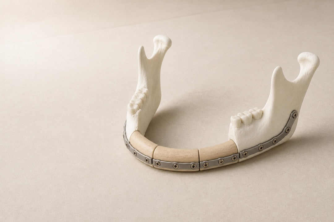
Head & Neck Reconstruction
Head & Neck Reconstruction
Reconstruction after cancer resection involving the tongue, oral cavity, pharynx, maxilla, or mandible, together with static reconstruction, nerve reconstruction, and muscle transfer for facial paralysis.
Read →
Extremity & Trunk Reconstruction
Sarcoma Reconstruction
Reconstruction of the extremities and trunk after sarcoma resection, preserving movement while minimizing donor-site burden.
Read →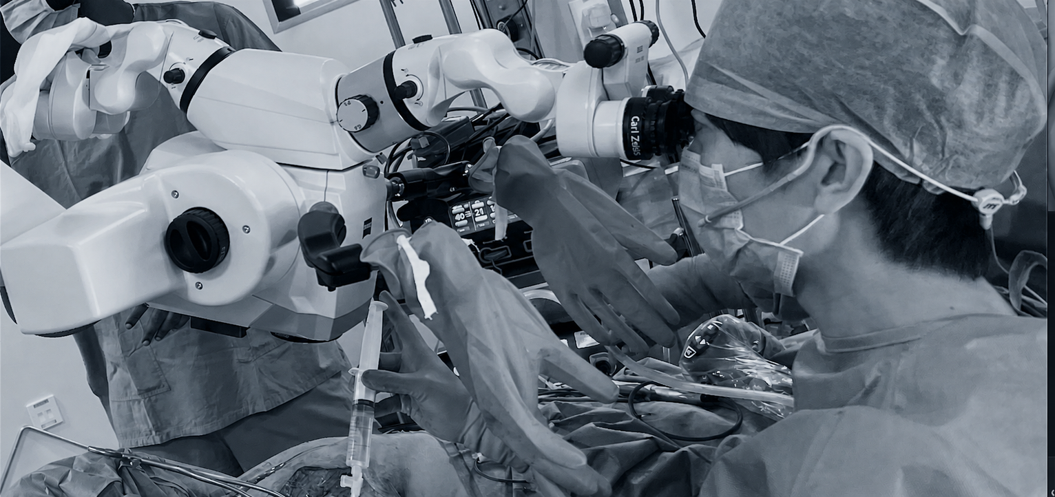
01
Breast Reconstruction
Breast Reconstruction
Breast reconstruction rebuilds a breast lost through cancer surgery. There are two principal approaches.
Using the Patient’s Own Tissue (Autologous Reconstruction)
Skin and fat are transferred with their blood supply from the abdomen or thigh to the breast, and the vessels are connected under the microscope. The reconstructed breast tends to feel soft, age naturally, and remain stable over time. The trade-offs are a longer operation and a scar at the donor site.
The donor site is selected together after considering body habitus, plans for pregnancy, previous operations, and patient preference.
Using an Implant
A tissue expander is used to stretch the skin before placement of a silicone implant. This avoids creating a new donor-site scar and shortens operating time. Suitability depends on factors including radiation therapy and skin condition.
What I Emphasize
Preoperative ultrasound is used to map the vessels and select the least invasive suitable flap. For example, when an SIEA flap can be used safely, I favor it over a DIEP flap. When exposing the internal mammary recipient vessels, my standard approach is to preserve the costal cartilage.
I also use intraoperative ICG fluorescence angiography to confirm perfusion of the transferred tissue rather than relying on appearance alone.
Neither approach is universally superior. The best option depends on age, the breast-cancer treatment plan, body habitus, work and lifestyle, and the patient’s priorities. During consultation, I explain the benefits and burdens of each with equal weight.
Liposuction and Fat Grafting After Reconstruction
After breast reconstruction, concerns about asymmetry, contour, or residual hollows are common. Fat grafting can refine the reconstructed side, while liposuction may help balance the opposite side.
This is the refinement stage of reconstruction. It requires the ability to read asymmetry in millimeters and place only the required volume in a plane where the graft can survive. This is precisely where reconstructive and aesthetic techniques overlap, and it is one reason I continue to practice both.
Breast reconstruction is not always completed in a single operation. The final result includes these refinements, which are considered from the initial surgical plan.
Nipple–Areola Reconstruction
Nipple–areola reconstruction may use local flaps to create nipple projection or graft part of the nipple–areola complex. Three-dimensional medical tattooing can also recreate visual depth. These procedures are provided at appropriate partner hospitals or clinics.
Liposuction, fat grafting, and three-dimensional medical tattooing are self-funded. The other breast-reconstruction treatments described here are covered by Japanese public health insurance.
02
Head & Neck Reconstruction
Head & Neck Reconstruction
After resection of cancer involving the tongue, oral cavity, pharynx, maxilla, or mandible, quality of life depends not only on appearance but also on the ability to eat, swallow, and speak. The reconstructive surgeon restores the defect during the same operation and aims to recover these functions as far as possible.
Soft-Tissue Reconstruction
In reconstruction of the tongue and oral mucosa, the thickness and shape of transferred tissue must be adjusted to the extent of resection and movement of the remaining structures. Some defects require substantial volume, while unnecessary bulk can impair oral movement. An appropriate flap is selected from the forearm, groin, thigh, or another site and shaped tosupport eating and speech.
Bony Reconstruction
When the maxilla or mandible is lost, vascularized bone such as fibula or iliac crest is transferred according to defect location and size. Mandibular reconstruction emphasizes contour and continuity; maxillary reconstruction restores separation between the oral and nasal cavities and their supporting framework. When useful, a 3D model is generated from CT data to plan bone cuts and positioning before surgery, considering facial form, occlusion, and future dental rehabilitation.
Facial Paralysis Reconstruction
For facial paralysis, treatment is selected according to its extent and duration. Options include static reconstruction to support the corner of the mouth, nerve repair or nerve transfer, and dynamic reconstruction with muscle transfer. The aims may include restoring eye closure, stabilizing the mouth, and recovering movement for smiling.
Secondary Problems After Reconstruction
Trismus, swallowing difficulty, and other problems may develop over time after surgery or radiotherapy. I also perform secondary procedures to address these issues.
03
Extremity & Trunk Reconstruction
Extremity & Trunk Reconstruction After Sarcoma Resection
In surgery for soft-tissue sarcoma or osteosarcoma, complete oncologic resection with an appropriate margin is the first priority. Skin, muscle, nerve, vessel, and bone may be removed together, leaving a wound that cannot be closed or a limb that cannot function. Reconstruction fills the defect during the same operation while preserving function where possible.
Minimizing Donor-Site Burden
This has been a particular focus of my work. Even in situations where a large muscle was traditionally transferred, preoperative ultrasound mapping of vascular anatomy can often allow reconstruction with a thin flap composed only of skin and fat. Preserving muscle also preserves strength at the donor site. The clinical effect of this approach has been reported in peer-reviewed publications.
Restoring Movement
The skin of the hands, feet, arms, and legs is thin, with tendon and bone immediately beneath it. Excessively bulky coverage can prevent wearing shoes or closing the hand. A thin flap is selected, and when muscle or tendon has been lost, it may be combined withtendon transfer—redirecting a remaining tendon to replace a lost function—so that reconstruction restores movement as well as coverage.
When Bone Is Lost
A bony defect may be reconstructed with vascularized fibula or iliac crest. Because the transferred bone remains living tissue, it can unite with surrounding bone and adapt to mechanical load over time.
Reconstruction After Repeated Recurrence
Sarcoma may recur locally. By a second or third operation, previously used recipient vessels may no longer be available and irradiated skin may be fibrotic. Reconstruction in these difficult settings requires maintaining several viable options.
Sarcoma care is coordinated with orthopedic oncology, medical oncology, and radiation oncology. The reconstructive plan is designed by the team once the extent of resection is established.
Technology
Visualize Before Operating
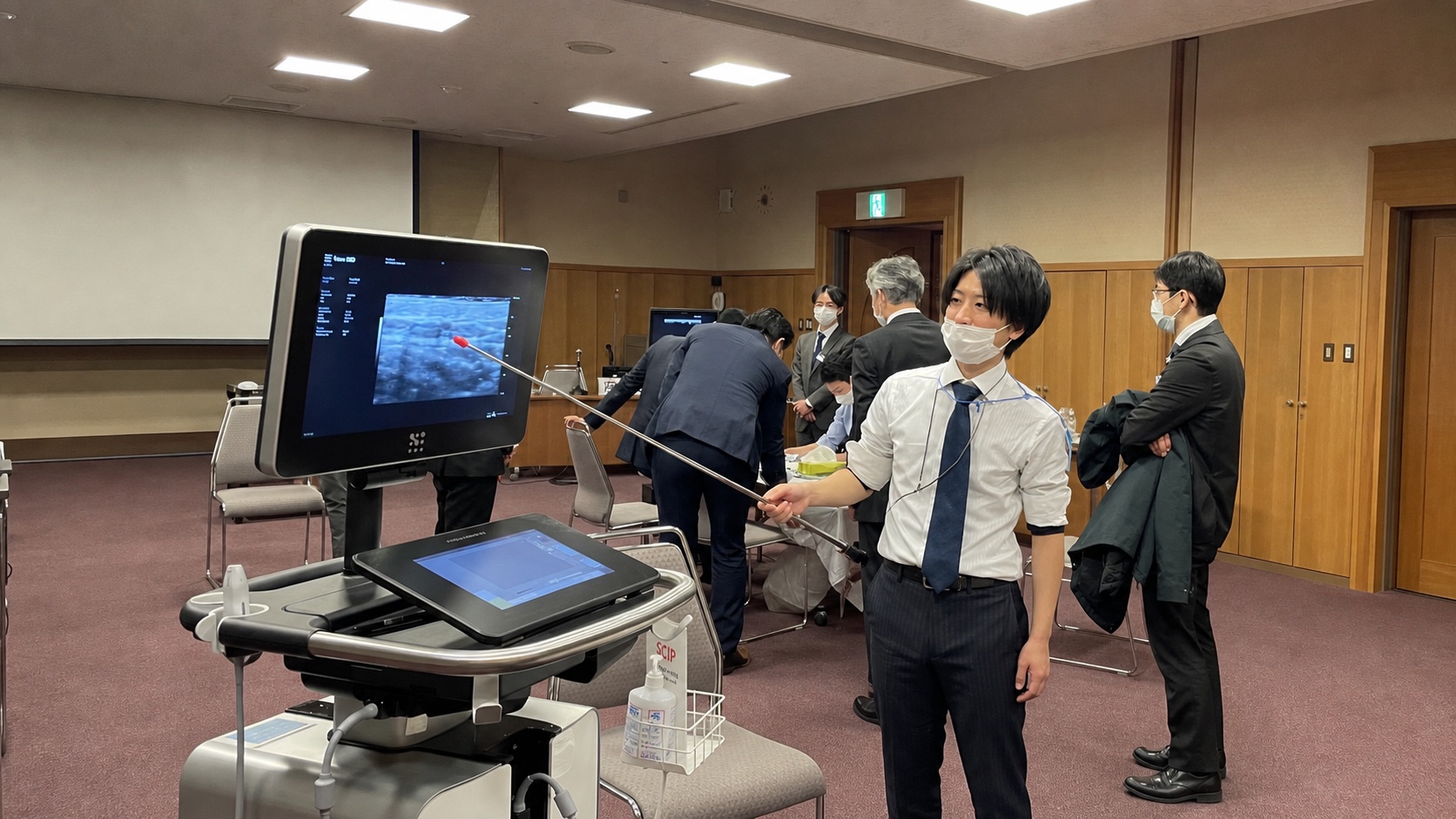
Teaching as an instructor at an ultra-high-frequency ultrasound course.
Ultra-High-Frequency Ultrasound
This technology provides much finer detail than conventional ultrasound and maps vessels smaller than 1 mm before surgery. Defining where to operate before incision can reduce dissection and operative time.
ICG Fluorescence Angiography
ICG fluorescence enables real-time intraoperative assessment of perfusion in transferred tissue and visualization of lymphatic flow, reducing reliance on assumption alone.
Operative Recording and Review
Operations are recorded and reviewed against outcomes. This archive informs publications and educational videos, and the lessons return to subsequent surgery.
These technologies and outcomes have been reported in peer-reviewed international journals. See the Academic for details.
Related Practice
Lymphedema Surgery
Removal of lymph nodes during cancer treatment can lead to swelling of an arm or leg—lymphedema. As part of reconstructive surgery, I perform lymphaticovenular anastomosis (LVA) and liposuction.
A separate page explains the treatment approach, perioperative pathway, limitations, and risks for patients who are scheduled for surgery and their families.